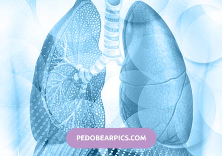
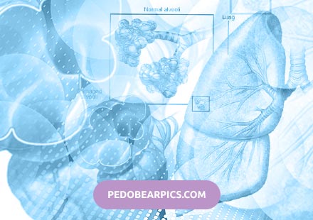
Prevention of lung disease
Disappointing statistics of today suggests that the most terrible lung diseases are the most common. Due to improper and inadequate nutrition, poor ecology and many bad habits of lung cancer and tuberculosis in recent years, came to the rank of government problems.
Talk about lung disease prevention It is difficult, because everyone knows that smoking is a harm to the body and a threat to the respiratory system. But at the same time, most people are extremely frivolous about this fact.
Quitting smoking is difficult, but in the name of health for such sacrifices worth making. In addition, we must not forget that passive smoking is no less dangerous than active. It is important to try to avoid places with high gas content, and at first symptoms of tuberculosis or other ills seek help from a doctor.

Prevention of respiratory diseases:
- For many smokers it is difficult to give up the bad habit. With a long smoking period, some people generally reject this possibility of prevention. But at the same time, as practice shows, the majority of former smokers, having abandoned the dangerous habit, no longer return to it;
- Spending time outdoors is useful. Leaving the city in the morning or on weekends, most people note the fact that it becomes much easier to breathe, constant fatigue and weakness of the body leave. Many doctors say that people who often go for walks in the woods or mountains suffer much less frequently. diseases respiratory organs;
- Proper nutrition is the basic rule of good health, good health and strong immunity. And the stronger the immune system in humans, the easier it is to cope with all diseases, including tuberculosis;
We warn tuberculosis
Effective tuberculosis prevention and other lung diseases primarily depends on how clean the air is constantly breathing people. According to medical statistics, often x-rays of the former smoker's lungs after 3 years show an identical picture with the lungs of a person who has never smoked before.
Also worth noting is the fact that any tuberculosis treatment or prevention of lung cancer should be carried out only after consulting a specialist. Do not self-medicate, do not experience fate and your own body, especially if you decide to use alternative medicine.
Dangerous complications of pulmonary tuberculosis
Tuberculosis is a common infectious disease with a tendency to a chronic relapsing course. This disease reflects a social condition, as it often develops in places where poverty, overcrowding, and a low level of education are prevalent. Contribute to the development of tuberculosis and the presence of some dangerous diseases: diabetes, AIDS, chronic lung diseases, alcoholism.
Forms of secondary tuberculosis
Tuberculosis in clinical and morphological forms are divided into primary, secondary and hematogenous.
Primary tuberculosis - the disease develops at the time of the first meeting with the pathogen - Koch's wand. Characteristic risk factors for this are childhood or weakened body defenses (for example, in HIV-infected people).
Hematogenous tuberculosis is said to occur when, after the initial infection, the infection spreads through the bloodstream. Any tissue and organs can be affected by tuberculosis.
The most common form of tuberculosis is secondary. The development of the disease occurs in adults, in whom primary tuberculous affect was formed and healed in childhood. Consensus about the source infections at the moment no. There are opinions that in case of secondary tuberculosis, the infection is reactivated from the healed old foci, and some experts believe that infection occurs by re-infection from the external environment.
Forms of secondary tuberculosis are many. Each proceeds with its own characteristics and characteristic complications.
- Acute focal pulmonary tuberculosis;
- Fibrous focal;
- Infiltrative;
- Tuberculosis;
- Acute cavernous;
- Fibro-cavernous;
- Caseous pneumonia;
- Cirrhotic tuberculosis.
Especially unfavorable in terms of the development of complications, the so-called "destructive" forms of tuberculosis, in which there is a breakdown of lung tissue, walls of the bronchi, vascular walls. These include cavernous, fibro-cavernous, cirrhotic tuberculosis, tuberculoma, caseous pneumonia.
Any form of tuberculous process can become destructive with late treatment.
What is dangerous pulmonary tuberculosis?
The disease itself is quite dangerous in that it has a long chronic course, adversely affecting the work of many organs and systems. And complications of pulmonary tuberculosis threaten the patient's life altogether. The most common complications consider in more detail.
Hemoptysis
Hemoptysis is a presence in the sputum of blood. The blood is excreted along with sputum when it is coughing, it can be in the form of veins or separated by spitting.
Hemoptysis is often found in destructive forms of pulmonary tuberculosis (fibrous-cavernous, cavernous, cirrhotic), when small vessels are damaged. A small amount of blood is released when you cough along with sputum. Over time, hemoptysis can develop into pulmonary hemorrhage when, as a result of the tuberculous process, the walls of larger vessels are damaged and blood is poured out of them.
Pulmonary hemorrhage
In cases where a significant amount of blood is secreted from the respiratory tract, pulmonary hemorrhage is indicated.
Bleeding is the discharge of blood in a volume in excess of 50 ml.
Pulmonary hemorrhage develops for the following reasons:
- Increased pressure in the pulmonary vessels;
- Increased vascular permeability;
- Arrosion of vessel walls in destructive forms of tuberculosis;
Features of pulmonary hemorrhage in pulmonary tuberculosis:
- Blood excretion occurs with cough tremors and characteristic "bubbling";
- Blood may be mixed with sputum or excreted in pure form;
- With pulmonary bleeding, the color of blood is bright red, the consistency is frothy.
If the volume of blood separated from the respiratory tract exceeds 500 ml, such bleeding is called profuse. This is a very dangerous condition in which death can quickly occur.
Spontaneous pneumothorax
Pulmonary tuberculosis may be complicated by the formation of spontaneous pneumothorax. Pneumothorax is a condition in which air accumulates in the pleural cavity. In the case of the tuberculous process, this occurs during the destruction of the lung tissue, as a result of which the pleural cavity communicates with the airways.
Spontaneous pneumothorax in the tuberculous process has its own characteristics:
- It develops slowly, the onset may be asymptomatic, pain increases as air enters the pleural cavity.
- Often complicated by the addition of a secondary infection with the development of pleural empyema (suppuration).
Requires surgical treatment (drainage of the pleural cavity) in combination with anti-tuberculosis (specific) and antibacterial therapy (non-specific).
Secondary amyloidosis
Amyloidosis is a pathological process by which the deposition of protein - amyloid in the tissues of organs. This is the result of a violation of metabolic processes in the body (in particular, protein metabolism), which inevitably develop during a long-term infection process, which is tuberculosis.
Most often this process affects the kidneys, spleen, liver, adrenal glands, walls of blood vessels.
Tuberculosis is a disease in which amyloidosis develops most frequently. This complication manifests itself in different ways, and it can be very difficult to diagnose amyloidosis in its initial stages. In most cases, the diagnosis of "amyloidosis" is established only after the death of the patient.
Observations show that kidney amyloidosis is most often diagnosed in tuberculosis patients. It manifests itself by edema, pronounced loss of protein in the urine, a decrease in the amount of urine released (oligouria). Death can occur from chronic renal failure.
Pulmonary heart formation
With a long-term tuberculous process, as well as with destructive forms of tuberculosis, changes in the cardiovascular system are observed, which are commonly called "pulmonary heart".
This concept includes the following changes by hearts and vessels:
- Hypertrophy of the right ventricle (due to increased pressure in the pulmonary circulation increases the load on the right ventricle, which leads to its hypertrophy and stretching).
- Violation of gas exchange (due to vascular lesions, lung tissue, chest deformity).
This leads to the fact that over time, patients with pulmonary tuberculosis become patients of cardiologists.
Symptoms of cardiopulmonary insufficiency can "hide" for a long time under the mask of the underlying disease. Dyspnea, cough, weakness can be regarded as symptoms of a tuberculous process in the lungs, and not problems with the heart muscle.
When examining such patients, one should pay attention to the discoloration of the skin, which occurs due to hypoxemia (oxygen starvation). Marked cyanosis of the skin and mucous membranes, which is called cyanosis. A change in coloration of the skin can occur only on certain parts of the body — the ears, the tip of the nose, the lips, the fingers and toes. In such cases, talking about acrocyanosis. In addition, with the development of heart failure, the appearance of pain behind the sternum, edema, choking cough are noted.
Spread of infection
Pulmonary tuberculosis can spread to other organs. The spread of infection can occur through Koch-infected sputum, which, standing out from the respiratory tract, enters the oral cavity, and can also be swallowed by the patient. This path is called companion.
So, against the background of pulmonary tuberculosis, bronchial tuberculosis, larynx, vocal cords, and tongue may develop.
The main principle of treatment of complications of tuberculosis is adequate therapy of the underlying disease. With effective anti-tuberculosis therapy, almost all complications can be reversed. Although in some cases, no surgical intervention can not do, as for example, with pneumothorax, pulmonary hemorrhage.
Candidiasis of the lungs: diagnosis and treatment
Candidiasis of the lungs is a fairly rare, but dangerous disease that immunocompromised people primarily face. The essence of this pathological process is that the lung tissue is affected by yeast-like fungi belonging to the genus Candida. As a rule, the infectious flora enters the respiratory system from a primary focus that is localized in the body. From a clinical point of view, most often this pathology is manifested by candidal pneumonia, but other options are possible. Timely initiated therapy allows for complete recovery in this condition. In advanced cases, it can cause severe respiratory failure and even subsequent death.
There is no exact information about the prevalence of lung candidiasis among the population. However, it has been reliably established that people of absolutely any age, including children, may encounter such a disease. Any dependence on gender is also not traceable. Interestingly, isolated lung disease is diagnosed much less frequently than generalized forms of candidal infection. Speaking of the generalized form, we mean the involvement in the pathological process of the skin, intestines, lungs and many other internal organs. A prerequisite for the development of this disease is a pronounced decrease in the level of immune protection. The level of mortality in this state can be from thirty to seventy percent, depending on what category sick people fall into.
As we have said, the causative agent candida lung is a conditionally pathogenic microorganism belonging to the genus Candida. It is a yeast-like fungus that normally is found in more than fifty percent of people. However, in the normal state of the body, this flora is not capable of causing him any harm. The situation changes with a decrease in the level of immune protection.
In most cases, fungal flora enters the respiratory system from other infectious foci. At the same time, depending on the mechanism of infection, the primary and secondary forms of this disease are distinguished. The primary form implies a cast into the lungs of a secret that contains the causative agent. In the secondary form, the fungal flora spreads with a current of lymph from the primary focus.

The most common lung candidiasis occurs in people suffering from various immunodeficiency states. An example of this is HIV infection. Other predisposing factors include various problems on the part of the endocrine system, existing inflammatory lesions in the lungs, bad habits, and malignant tumors. Often, a decrease in the level of immune protection is due to the long-term administration of drugs that have a suppressive effect on it. These include glucocorticosteroids and cytotoxic drugs.
Primarily under the influence of the fungal flora, small inflammatory foci are formed in the lung parenchyma, the central part of which is necrotized. Quite often small bronchi are involved in the pathological process. Often, pulmonary candidiasis is complicated by the formation of purulent cavities and cavities, and then granulation-fibrous changes. In general, certain mechanisms may prevail in each particular patient.
Symptoms characteristic of pulmonary candidiasis
As we have said, most often this disease occurs in the form of candidal pneumonia. Symptoms may be acute in this case, however, most often such pathology acquires a protracted course with the occurrence of periodic exacerbations. Initially, a sick person begins to complain of bouts of dry cough, which may be accompanied by a discharge of a very small amount of sputum. There is a mandatory general intoxication syndrome, including subfebrile or febrile fever, weakness and malaise. Other specific manifestations include shortness of breath and pain in chest, aggravated by breathing.
Often, the inflammatory process in the lungs, caused by exposure to fungal flora, is complicated by symptoms indicating pleurisy. At the same time in the pleural cavity a sufficiently large amount of exudate, having a serious or hemorrhagic character, is formed. There is another clinical variant of this disease - miliary. It is characterized by the appearance of a painful cough, in which a small amount of mucous- bloody sputum is excreted. Additionally, noted the presence of difficulty exhaling.
Sometimes candidal lesion of the lungs occurs in a latent form, especially in people who are on artificial lung ventilation. Very often associated symptoms resemble other chronic lung diseases, which creates certain difficulties in diagnosis. The most severe forms of this pathological process are usually diagnosed in young children.
Diagnosis and treatment of the disease
First of all, it is possible to suspect specific changes in the lungs with the help of an auscultatory study. Mandatory radiography of the lungs. In doubtful cases, computed tomography is used. In addition, this disease is an indication for the appointment of microscopic and culturalexamination of sputum. Detect the causative agent is also possible with the help of various serological tests, but they are not the basis for diagnosis.
Treatment for pulmonary candidiasis consists of the use of specific antifungal drugs. In this case, antimycotics are used both systemically and in the form of inhalation. Additionally, prescribed vitamin therapy, detoxification measures, expectorant drugs and so on.
Prevention of pulmonary candidiasis
The main method prophylaxis is an increase in the level of immune protection. To this end, you should try to adhere to a healthy lifestyle. In addition, it is recommended to promptly eliminate problems from the endocrine system and engage in the treatment of bronchopulmonary diseases.
Prevention of pulmonary candidiasis
The main method prophylaxis is an increase in the level of immune protection. To this end, you should try to adhere to a healthy lifestyle. Besides,
5 symptoms that indicate problems in the lungs
Lungs - this paired organ plays a very important role in the human body, providing gas exchange between the atmosphere and blood. As for the latter, about 9% of the total volume of this fluid mobile connective tissue of the internal environment is concentrated in the lungs. The lungs contain about 450 ml of blood, but the nerve endings are completely absent in this organ, and therefore they cannot signal any malfunctions in their work. Therefore, it is important to pay attention to symptoms that directly or indirectly may indicate respiratory diseases.
Weight Going: when standing appointment with the doctor on reception?
Unreasonable weight gain or weight loss may indicate problems in the lungs. In the first case, we can talk about pulmonary edema, indicating the accumulation of fluid in the respiratory system. In an ordinary person, when breathing in, the lungs are filled with air, and in a patient - with fluid. This process is dangerous because of its suddenness, because acute pulmonary edema can lead to serious irreversible consequences. In any case, when the first symptoms associated with sudden shortness of breath, wheezing, the appearance of weakness and dizziness, pink frothy saliva is necessary to ring as soon as possible by phone and call an ambulance.
The same signs point to chronic pulmonary edema, but they develop gradually. Weight gain may indicate congestive heart failure, so do not pull, but an urgent need to make an appointment with a doctor. Weight loss, if a person has not changed his eating habits, may indicate chronic obstructive pulmonary disease. The fact is that with this disease the lungs greatly increase in size, the distance between them and the stomach decreases, and with a hearty meal, there is pressure, discomfort, which causes a person to reduce the amount of food consumed. Foods high in fiber, spicy, salty and carbonated foods only worsen the patient's condition.
Edema - a warning sign
Swelling of the ankles and feet at least once in my life, every person has come across. If this happens only occasionally and is explained by nutritional errors and other lifestyle features, such as hiking, then there is no cause for concern. Another thing, if this is observed constantly. In the human body, everything is interconnected, and swelling of the legs may indicate problems in the work of the respiratory system. Any disruption of the lungs causes oxygen deficiency, which primarily affects the circulatory and lymphatic systems. The circulation of fluids slows down, the internal organs, including the kidneys, are not supplied with blood as expected, and therefore cannot work effectively and perform their task of removing excess fluid from the body, which leads to edema.
Do not leave such a symptom without attention. You need to call the caller confidence in the clinic and sign up to doctor on reception. Even if a specialist eliminates lung disease, you need to get to the truth and determine the exact cause of edema. It does not matter whether it is venous insufficiency, lymphedema or thrombosis, the pathology must be treated to avoid a variety of unpleasant consequences.
Dyspnea is another dangerous symptom
It is quite natural that breathing gets off and quickens during physical exertion and sports, but it is quite another thing when similar symptoms occur on level ground. The heart and lungs are the two main organs involved in the transport of oxygen to cells and tissues and the elimination of carbon dioxide, which means that any disruption in their work will invariably affect the quality of respiration. The fears of specialists cause both sudden, acute dyspnea, and gradually developing changes in the rhythm and depth of breathing.
If a person notices that even at rest, especially in the horizontal position, there are problems with breathing, feeling short of breath, it is a chance to phone the hospital and sign up to doctor on reception. It happens so, that difficulties arose ayut only inhale or exhale. In the latter case, this is characteristic of the narrowing of the lumen of the small bronchi and bronchioles, which develops during bronchial asthma. Chronic dyspnea may be a consequence of the already mentioned chronic obstructive pulmonary disease, interstitial disease, etc.
Chronic weakness and fatigue
The lack of oxygen leads to a decrease in the level of hemoglobin and the number of red blood cells, and this negatively affects the body's ability to produce energy to maintain its vital activity. When the lungs malfunction and oxygen deficiency, the person suffers from constant weakness, and fatigue becomes his usual companion. Such symptoms are most often experienced by people with pulmonary fibrosis. They describe their condition as relentless exhaustion, which seriously impairs their quality of life.
Pulmonary fibrosis is an autoimmune disease, the causes of which and the numerous symptoms are still to be studied by specialists. Why it causes fatigue remains to be seen, but scientists believe that the whole thing is fast, shallow breathing during sleep. Thus, the body tries to compensate for the lack of oxygen, but it reduces the duration of deep sleep, which means that a person wakes up not rested enough and feels lethargy and drowsiness even at the beginning of the day.
What diseases will tell a persistent cough?
If the swelling of the legs can be the result of a variety of health problems, then cough directly indicates a malfunction of the respiratory system. However, not everyone pays attention to this alarming symptom. Cough is the main symptom of colds and flu, but many suffer from these diseases on their feet, not considering them so serious as to pay much attention to treatment. They begin to worry only when the cough is delayed by more than 2 weeks. But such a reaction of the organism in the form of forced expiration through the mouth can really be the result of not only a banal respiratory infection.
Lesions of the pulmonary vessels and lung tissue, malformations, allergies, asthma, mycosis - all of these diseases may be accompanied by coughing and require more serious treatment. If it does not pass within 4-8 weeks, reduces the quality of sleep and provokes constant chronic fatigue, then this is a reason to consult with a specialist.
Diagnosis of diseases of the bronchi: the main methods
How to diagnose bronchitis
The inflammatory process that develops in the bronchi during an infectious process is called bronchitis. He is diagnosed during auscultation (listening to the chest with the help of a phonendoscope) and ascertaining the characteristic deep moist rales. In addition, a symptom of bronchitis is a characteristic wet cough.
However, all the above diagnostic criteria do not always correspond exactly to the disease. For example, asthma, tuberculosis, cancer and other pathologies may be hidden under the guise of bronchitis.
TO Unfortunately, even experts do not always think about it, and when they hear characteristic wheezing, they prescribe a handful of pills. AND only with their absolute uselessness direct the patient to more serious research, while losing time.
BUT what should be done? AT First of all, take a sputum sample. This is done as follows. From the bronchi take a sample of sputum and make it seeding on a Petri dish. Then, after a few days, they are studying what, in fact, has grown on them (which microorganisms colonize the patient's bronchi). And, if pathogens are found, then appropriate treatment is prescribed (antibiotics, anti-inflammatory, mucolytics, expectorant - with viscous sputum).
BUT if it is not at infections, or not only in it, it is necessary to appoint a chest x-ray, and, without fail, bronchoscopy.
What is bronchoscopy?
Bronchoscopy is the study of the condition of the bronchi with the help of special optical systems - bronchoscopes. They are flexible or rigid optical fibers to which a light source is connected. Bronhosokop entered through the trachea in the bronchi. BUT The "picture" of what is happening is visualized through an external eyepiece. Thanks to the light beam through the fiber, you can see the condition of the bronchi and their mucous membranes, see the inflammatory process, the contents of the bronchi, the presence of tumors in the bronchi or their compression by lymph nodes. In inflammatory processes, bronchoscopy reveals a change in the normal color of the bronchi: they become reddish, and the shell is edematous, its vascular pattern changes.
In atrophic processes, the mucous membrane becomes thinner, and the bronchi themselves look "gaping."
Bronchoscopy capabilities
Bronchoscopy allows you to diagnose purulent bronchitis, bronchiectasis, tuberculosis and other lung pathologies, neoplasms, systemic lung diseases. Using this method, it is possible to determine the sources of bleeding in the bronchopulmonary. system.
WITH using bronchoscopy, you can perform a diagnostic biopsy of the tissues of the bronchi, lungs and lymph nodes, and also - lavage (washing) of small bronchi with the purpose of studying the composition of lavage waters. Through a bronchoscope, special sensors are analyzed, analyzing the composition of exhaled air, and medicines - for purulent bronchitis, for example, or for abscesses.
Also, with the help of a bronchoscope, foreign bodies can be extracted from the bronchi, or bronchial drainage from accumulated sputum can be performed.
By the way, on the basis of bronchoscopy, operations were invented that allow penetration into the cavities of the bronchi and lungs without surgical opening of the chest cavity.
Thus, bronchoscopy is a multifunctional method by which it is possible not only to diagnose the disease, but and if necessary, local treatment of existing pathologies.
Preparation for bronchoscopy
Bronchoscopy is a rather difficult procedure for the patient, therefore, before it is performed, premedication is performed - prior introduction of medications with bronchodilator and anesthetic (anesthetic) effect. In addition, the patient must be psychologically prepared for such a complex ordeal.

It should be remembered that in some cases, bronchoscopy should be used with caution or do is to abandon its holding!
TO Such cases include: bronchial asthma, coronary heart disease, diseases of the blood coagulation system.
AT In these cases, it is better to have a CT scan or magnetic resonance imaging.
Named the two most common lung diseases
University of Washington experts estimate that the two most common lung diseases in the year before last claimed the lives of 3.6 million people on the planet.
During the study, scientists analyzed data from 1990 to 2015, collected in 188 countries. According to statistics, in 2015, 3.2 million people died due to chronic obstructive pulmonary disease (COPD), and 400 thousand - due to asthma. And asthma occurs 2 times more often than COPD, but the latter is 8 times more deadly. It should be added that, according to WHO, in 2015, COPD was the fourth in the ranking of causes of death for people - after heart disease (9 million), stroke (6 million) and lower respiratory tract infections (slightly more than 3.2 million).
The prevalence of asthma has increased over the years by 13%, but the number of deaths has declined by more than a quarter. At the same time, the prevalence and mortality rates of COPD have decreased over this time, but deaths have increased by 12%. Scientists explain these statistical paradoxes by population growth - against the background of the development of medicine and technology.
According to scientists, these diseases attract less attention than such sadly "popular" ailments as cardiovascular diseases, cancer or diabetes. The data they received is a kind of reminder that efforts should be redirected to a great extent to combat them.
Symptoms for cavernous pulmonary tuberculosis
Cavernous pulmonary tuberculosis is a rather difficult pathological process, which, as a rule, has a secondary nature. This disease is characterized by destructive changes in the lung tissue, resulting in the formation of isolated thin-walled cavities, called caverns. At the same time in the tissues surrounding the cavity, there is no pronounced perifocal inflammation and fibrous changes. This pathology with the necessary treatment has a favorable prognosis. However, in some cases, it leads to the development of serious complications, for example, pulmonary hemorrhage, respiratory failure and so on.
As we have said, in the overwhelming majority of cases, cavernous pulmonary tuberculosis is the result of some other form of this disease. Most often it is preceded by an infiltrative form, much less often - disseminated or focal. In the event that the treatment of this pathology was not carried out, there is a high probability of the occurrence of fibrous changes in the cavity walls, which indicates its transition to the fibro-cavernous form.
Pathological process occurring in the cavernous form is detected in about five percent of people with tuberculosis. Most often, adults encounter it. Among children, this disease is detected less frequently.
The causative agent of this disease is a specific bacterium called Mycobacterium tuberculosis. It is motionless and has a rod-shaped form. The important point is that this bacterium is fairly well preserved under the influence of environmental factors. High and low temperatures, chemical disinfectants do not cause her death immediately. The most sensitive M. tuberculosis to direct ultraviolet rays.
Most often, with this pathological process, an air-borne mechanism of infection is implemented. In other words, an infected person carrying the disease in an open form releases a pathogen into the environment with particles of its saliva. Much less often the transmission of infection is caused by the alimentary, contact-household or transplacental routes.
The important point is that when M. tuberculosis penetrates the human body, tuberculosis does not always develop. Such factors as immunodeficiency states, bad habits, unfavorable social situation, existing oncological pathologies and much more are of great importance in its occurrence.
The mechanism of development of the cavernous form of this disease lies in the fact that a cavity filled with necrotic tissues forms in the initially formed inflammatory focus. In this case, the necrotic tissue is represented by caseous masses. After some time, there is a gradual rejection of the caseous masses through the lumen of the draining bronchus. In their place remains a cavity in which air or liquid is subsequently detected.
The classification of cavernous pulmonary tuberculosis includes several of its varieties: fresh disintegrating, fresh elastic, encapsulated, fibrous, and sanitized.
With fresh decaying species, destructive cavities are formed in the lungs, which are not yet separated from the surrounding tissues. A fresh elastic variety is established if the inner and middle shells have been formed, which delimit the cavity from the surrounding tissues and consist of a layer of caseous masses and granulation tissue. The encapsulated variety is also characterized by the appearance of an external connective tissue capsule. The fibrous type implies the launch of fibrous changes in the wall of the cavernous cavity. In a sanitized variety, granulation and caseous masses are rejected from the cavity.
Depending on the size of the arisen defect, small, medium and large cavities are distinguished. The diameter of the small cavities does not exceed two centimeters, and medium - five centimeters. Large caverns occur when their diameter is more than five centimeters.
Symptoms for cavernous tuberculosis
Most often, such a pathological process is one-sided. As a rule, specific symptoms appear three or four months after the ineffective treatment of other forms.
The brightest clinical manifestations are present in the phase when the cavernous cavity is just being formed. A sick person complains of a paroxysmal cough that is productive. In some cases, blood impurities are detected in the sputum. Quite often at this stage there are symptoms indicating general intoxication of the body. These include fever, severe weakness, and so on. During auscultation, you can listen to heavy wheezing, having a wet nature.
The clinical picture after cavity formation is expressed much less intensely. It is manifested by such symptoms as a periodic rise in temperature to subfebrile values, constant malaise, loss of appetite, weight loss, and so on.
Diagnosis and treatment of the disease
This disease is diagnosed on the basis of a concomitant clinical picture, anamnesis, as well as additional research methods. As we have said, most likely in the patient's history there will be other forms of tuberculosis infection. Additional methods include radiography of the lungs and bronchoscopy. Mandatory analysis of sputum for M. tuberculosis.
The first treatment of cavernous pulmonary tuberculosis involves the appointment of specific anti-tuberculosis therapy, which is used according to a specially designed scheme. With the ineffectiveness of the treatment may require surgery.
Prevention of cavernous tuberculosis
The main method of prevention is the correct and timely treatment of other forms of tuberculosis infection.